Uterus

1. Introduction 100%
Role of Ultrasound in Gynecology
Common Indications: AUB, Pain, Infertility, Mass Evaluation
Transabdominal vs Transvaginal Approaches
Advantages and Limitations
2. Uterine Anatomy 100%
Fundus, Body, Isthmus, and Cervix
Endometrial Cavity, Myometrium, Serosa
Junctional Zone (Adenomyosis Marker)
Uterine Arteries and Vascular Supply
Position (Anteverted, Retroverted, Axial)
3. Scanning Techniques 100%
Transvaginal (TVS): High-Resolution, Empty Bladder
Transabdominal (TAS): Full Bladder for Overview
Sagittal and Transverse Planes
Use of Doppler and 3D Ultrasound
Measuring Uterus and Endometrial Thickness
4. Normal Uterus Appearance 100%
Age-Dependent Uterine Size and Shape
• Endometrial thickness
• Uterine volume
• Uterine wall thickness (UWT)
• LSCS Scar thickness
• Cervical length
• RMT
• Measurement of internal indentation angle
Endometrial Echo: Triple Line, Homogenous, Thickness per Phase
Myometrium: Homogeneous and Isoechoic
Junctional Zone: Intact, Smooth, Hypoechoic
5. Congenital Uterine Anomalies 80%
1. MRKHS/Müllerian duct anomaly
Müllerian duct anomaly -Class-i (Uterine agenesis/uterine hypoplasia)
• Class-i-a Vaginal hypoplasia
• Class-i-b Cervical hypoplasia
• Class-i-c Fundal hypoplasia
• Class-i-d Tubal hypoplasia
• Class-i-e combined hypoplasia
Müllerian duct anomaly -Class-ii (Unicornuate Uterus)
• Class-ii-a Unicornuate Uterus (communicating horn)
• Class-ii-b Unicornuate Uterus (Non-communicating horn)
• Class-ii-c Unicornuate Uterus (No cavity)
• Class-ii-d Unicornuate Uterus (No horn)
• Müllerian duct anomaly -Class-iii
Müllerian duct anomaly -Class-iv- (Bicornuate uterus).
• Class-iva (Bicornuate bicollis)
• Class-ivb (Bicornuate unicollis)
• Class-v (septate uterus).
• Class-v-a (Partial/subseptate uterus)
• Class-v-b (Complete septate uterus)
• Class-vi (Arcuate uterus)
2. DES-related T-shaped Uterus
3. Role of 3D US, MRI for Confirmation
6. Myometrial Pathologies 100%
1. Fibroids (Leiomyomas)
2. Adenomyosis
Diffuse Adenomyosis
Focal Adenomyosis
Cystic Myometrial Cysts and Striated Pattern
Thickened Junctional Zone >12 mm
7. Endometrial Disorders 100%
Endometrial Hyperplasia
Endometrial Polyp
Endometritis (Acute/Chronic)
Endometrial Carcinoma
Saline Infusion Sonography (SIS) for Cavity Evaluation,br.
Others common findings
Bulky uterus
Nabothian cyst
Cervicitis
Hematometra
Pyometra
Hematocolpos
Vaginal cyst
Gartner duct cyst
8. Vascular and Functional Disorders 30%
Abnormal Uterine Bleeding (AUB) Evaluation
Arteriovenous Malformation (AVM)
Uterine Synechiae (Asherman’s Syndrome)
Menstrual Phase Correlation with Endometrium
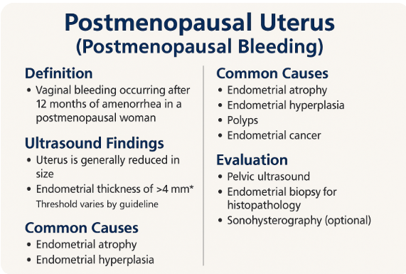
9. Postmenopausal Uterus 0%
Normal Involutional Changes
Endometrial Stripe < 5 mm (Normal)
Postmenopausal Bleeding Evaluation
Endometrial Thickness Guidelines and Biopsy Indications
10. Interventional and Follow-up Role 0%
Hysterosonography (SIS)
US-Guided Fibroid Biopsy
Post-Myomectomy or Hysteroscopic Resection
Treatment Monitoring (GnRH, IUD, Ablation)
11. Case Studies and Quiz Section 0%
Fibroid Localization and Classification
Endometrial Carcinoma vs Hyperplasia Cases
Uterine AVM Diagnosis Challenge
Interactive MCQs with Image Interpretation

CEUS for Renal Masses & Perfusion

CEUS for Renal Masses & Perfusion 0%
1. Introduction to Renal CEUS
Role of CEUS in Renal Imaging
Safety and Non-nephrotoxic Nature of Microbubble Contrast
Indications: Renal Mass Characterization, Infarcts, Perfusion Defects
2. CEUS Protocol for Kidney
Contrast Agent (e.g., SonoVue, Lumason)
Patient Prep and Injection Timing
CEUS Phases: Corticomedullary (10–30 sec), Nephrographic (30–120 sec), Late Phase (>120 sec)
Use of Dual Imaging Mode (B-mode + Contrast)
3. Renal Mass Evaluation
Cystic Lesions: Bosniak Classification with CEUS
Simple vs Complex Cysts
Solid Renal Masses (RCC, Oncocytoma, Angiomyolipoma)
Enhancement Patterns and Washout in Malignancy
4. CEUS in Renal Perfusion
Renal Infarction: Wedge-shaped Non-enhancement
Acute Cortical Necrosis
Pyelonephritis and Focal Nephritis
Transplant Kidney Perfusion Assessment
5. Comparison with CT/MRI
CEUS vs CT in Cystic Lesion Classification
CEUS in Patients with Renal Insufficiency
Role in Lesions Indeterminate on CT
CEUS as Follow-up Modality Post-Biopsy/Ablation
6. Pitfalls and Limitations
Pseudo-enhancement in Deep Lesions
Artifacts and Motion Sensitivity
Small Cortical Lesions May Be Missed
Differentiation Between RCC Subtypes Remains Limited
7. CEUS in Renal Intervention
Guidance for Biopsy of Indeterminate Masses
Assessment of Post-ablation Viability
Follow-up for Treated Tumors
Monitoring Transplant Complications
8. Case Studies and Quiz Section
CEUS in Complex Renal Cysts (Bosniak IIF–IV)
Differentiation of RCC vs AML
Real-Time CEUS Flow Interpretation
MCQs and Image-Based Diagnostic Challenges

Renal Dromedary hump

117
Case Study
Renal Dromedary hump
Dromedary hump is a normal anatomical variant of the kidney characterized by a localized bulge on the lateral border of the left kidney, typically due to impression from the adjacent spleen. It mimics a mass but maintains the normal renal cortical echotexture and vascularity on imaging, distinguishing it from pathological lesions.

Patient presents for routine abdominal ultrasound. No specific urinary symptoms. No history of flank pain, hematuria, fever, or weight loss. No known renal pathology. Incidental finding during evaluation.

Findings

image
📄 Report Sample Line- Dromedary hump
The left kidney shows a localized cortical bulge along its lateral border, maintaining normal renal cortical echotexture and vascularity, consistent with a Dromedary hump — a normal anatomical variant.

Conclusion

📋 Localized cortical bulge on the lateral aspect of the left kidney represents a Dromedary hump, a normal anatomical variant with no pathological significance.

Recommendation:


Dromedary Hump - Kidney

Causes

  • External impression of the spleen on the developing left kidney during fetal life
  • Results in a focal bulge on the lateral border of the kidney
  • Considered a normal anatomical variant, not a disease

Symptoms

  • Usually asymptomatic and discovered incidentally
  • No pain, urinary symptoms, or hematuria
  • Does not impair kidney function

Diagnosis

  • Ultrasound (USG): Shows a cortical bulge with uniform echotexture and normal Doppler flow
  • CT or MRI: Used for confirmation if needed; helps rule out renal mass
  • Key Feature: Maintains normal renal parenchyma appearance, unlike tumors


1. What is a Dromedary hump in renal ultrasound?
A. A renal artery aneurysm
B. A pathological renal mass
C. A normal cortical bulge of the kidney
D. A cyst in the renal pelvis
👉 Explanation: A Dromedary hump is a benign cortical bulge, often mistaken for a mass but part of normal kidney anatomy.

2. On which kidney is a Dromedary hump most commonly seen?
A. Right kidney
B. Left kidney
C. Both kidneys equally
D. Neither kidney
👉 Explanation: It is more common on the left kidney due to pressure from the adjacent spleen.

3. What typically causes a Dromedary hump?
A. Renal trauma
B. Liver compression
C. Splenic impression during development
D. Kidney stone
👉 Explanation: The spleen presses against the developing kidney, creating a persistent bulge.

4. What does a Dromedary hump look like on ultrasound?
A. Anechoic fluid-filled lesion
B. Hyperechoic with shadowing
C. Isoechoic with the renal cortex
D. Heterogeneous with calcifications
👉 Explanation: It appears isoechoic to surrounding renal cortex and blends in with normal tissue.

5. What is the clinical importance of a Dromedary hump?
A. It requires surgery
B. It is a sign of cancer
C. It is a normal variant with no clinical significance
D. It causes urinary tract obstruction
👉 Explanation: Dromedary humps are benign and do not require treatment.

6. How is a Dromedary hump usually discovered?
A. During surgery
B. Through physical examination
C. Incidentally on imaging
D. With biopsy
👉 Explanation: It is usually found incidentally during abdominal imaging.

7. What imaging modality is most useful in identifying a Dromedary hump?
A. X-ray
B. IVP
C. Ultrasound
D. PET scan
👉 Explanation: Ultrasound is typically used to identify renal cortical structures like Dromedary humps.

8. How can you differentiate a Dromedary hump from a renal tumor?
A. Tumor has cystic areas
B. Tumor shows necrosis
C. Dromedary hump maintains normal echotexture and blood flow
D. Tumor is located only in lower pole
👉 Explanation: Unlike tumors, Dromedary humps have uniform echotexture and normal Doppler flow.

9. What is the next step if there’s doubt between a tumor and a Dromedary hump?
A. Immediate surgery
B. Start antibiotics
C. Further imaging like CT or MRI
D. Ignore and observe
👉 Explanation: CT or MRI can provide more detailed images to rule out masses.

10. In which age group is a Dromedary hump most commonly found?
A. Children
B. Elderly
C. Any age
D. Neonates only
👉 Explanation: It can occur in any age group and is developmental in origin.

11. What part of the kidney is usually involved in a Dromedary hump?
A. Renal pelvis
B. Upper lateral cortex
C. Medulla
D. Lower pole
👉 Explanation: It typically affects the upper lateral border of the left kidney.

12. What is the treatment for a confirmed Dromedary hump?
A. Surgical removal
B. Biopsy
C. No treatment is required
D. Radiation
👉 Explanation: No treatment is needed for a benign Dromedary hump.

13. What role does Doppler ultrasound play in evaluating a Dromedary hump?
A. Measures kidney size
B. Detects stones
C. Confirms normal vascularity
D. Detects infections
👉 Explanation: Doppler shows normal blood flow in a Dromedary hump, unlike tumors.

14. Which condition can a Dromedary hump be mistaken for?
A. Pyelonephritis
B. Nephrolithiasis
C. Renal tumor
D. Hydronephrosis
👉 Explanation: It can mimic renal tumors on imaging due to its bulging appearance.

15. What confirms the diagnosis of a Dromedary hump on ultrasound?
A. Irregular shape
B. Central calcification
C. Continuity with renal cortex and normal blood flow
D. Mass effect on collecting system
👉 Explanation: A hump that shows same echotexture as cortex and normal Doppler flow confirms it's benign.


1. रेनल ड्रोमेडरी हंप क्या है?
A. रेनल धमनी का एन्यूरिज्म
B. एक रोगजनक गुर्दा ट्यूमर
C. गुर्दे की सामान्य कॉर्टिकल उभार
D. रेनल पेल्विस की सिस्ट
👉 व्याख्या: Dromedary hump एक सामान्य शारीरिक भिन्नता है, जो गुर्दे की सतह पर एक उभार के रूप में दिखाई देती है।

2. Dromedary hump सामान्यतः किस गुर्दे में पाया जाता है?
A. दायां गुर्दा
B. बायां गुर्दा
C. दोनों गुर्दों में
D. किसी में नहीं
👉 व्याख्या: यह अधिकतर बाएं गुर्दे में देखा जाता है क्योंकि उस पर प्लीहा का दबाव पड़ता है।

3. Dromedary hump बनने का कारण क्या है?
A. गुर्दे पर चोट
B. यकृत का दबाव
C. भ्रूण अवस्था में प्लीहा द्वारा दबाव
D. मूत्रमार्ग में रुकावट
👉 व्याख्या: भ्रूण विकास के समय प्लीहा द्वारा गुर्दे पर पड़ने वाले दबाव से यह उभार बनता है।

4. अल्ट्रासाउंड में Dromedary hump कैसा दिखता है?
A. ऐनीकोइक द्रव से भरा
B. छायायुक्त हाइपरइकोइक
C. सामान्य गुर्दा कॉर्टेक्स के समान इकोइक
D. मिश्रित और अनियमित
👉 व्याख्या: यह सामान्य गुर्दा टिशू की तरह ही इकोटेक्सचर दिखाता है, जिससे यह सौम्य लगता है।

5. Dromedary hump का चिकित्सीय महत्व क्या है?
A. तत्काल सर्जरी की आवश्यकता
B. यह कैंसर हो सकता है
C. यह एक सामान्य शारीरिक भिन्नता है
D. यह पेशाब में रुकावट करता है
👉 व्याख्या: Dromedary hump हानिरहित होता है और इसका इलाज आवश्यक नहीं होता।

6. Dromedary hump का पता कैसे चलता है?
A. ऑपरेशन के दौरान
B. शारीरिक परीक्षण से
C. इमेजिंग (जैसे अल्ट्रासाउंड) के दौरान
D. बायोप्सी से
👉 व्याख्या: यह सामान्यतः अल्ट्रासाउंड के दौरान संयोगवश पाया जाता है।

7. Dromedary hump की पहचान के लिए कौन सी इमेजिंग सर्वोत्तम है?
A. एक्स-रे
B. IVP
C. अल्ट्रासाउंड
D. PET स्कैन
👉 व्याख्या: अल्ट्रासाउंड गुर्दे की सतह की संरचनाओं को पहचानने के लिए सर्वोत्तम है।

8. Dromedary hump को ट्यूमर से कैसे अलग करें?
A. ट्यूमर में सिस्ट होते हैं
B. ट्यूमर में मृत ऊतक होता है
C. Dromedary hump में सामान्य इकोटेक्सचर और रक्त प्रवाह होता है
D. ट्यूमर हमेशा निचले ध्रुव में होता है
👉 व्याख्या: Dromedary hump में डॉपलर से सामान्य रक्त प्रवाह दिखता है और यह गुर्दे की सतह से जुड़ा होता है।

9. यदि संदेह हो कि यह ट्यूमर है, तो अगला कदम क्या होगा?
A. तत्काल सर्जरी
B. एंटीबायोटिक देना
C. CT या MRI द्वारा पुष्टि
D. इसे अनदेखा करें
👉 व्याख्या: CT/MRI जैसी इमेजिंग से ट्यूमर और हंप में अंतर किया जा सकता है।

10. Dromedary hump किस आयु वर्ग में देखा जा सकता है?
A. केवल बच्चों में
B. वृद्धों में
C. किसी भी उम्र में
D. केवल नवजातों में
👉 व्याख्या: यह किसी भी उम्र के व्यक्ति में पाया जा सकता है क्योंकि यह एक विकासात्मक विशेषता है।

11. गुर्दे का कौन सा भाग Dromedary hump से प्रभावित होता है?
A. पेल्विस
B. ऊपरी बाहरी कॉर्टेक्स
C. मेडुला
D. निचला ध्रुव
👉 व्याख्या: यह आमतौर पर बाएं गुर्दे के ऊपरी बाहरी भाग पर होता है।

12. पुष्ट Dromedary hump के लिए इलाज क्या है?
A. सर्जिकल हटाना
B. बायोप्सी
C. कोई इलाज नहीं चाहिए
D. रेडिएशन थेरेपी
👉 व्याख्या: पुष्ट Dromedary hump के लिए कोई इलाज आवश्यक नहीं है क्योंकि यह हानिरहित होता है।

13. डॉपलर अल्ट्रासाउंड Dromedary hump में किसलिए उपयोगी है?
A. गुर्दे का आकार मापने के लिए
B. पथरी की पहचान के लिए
C. सामान्य रक्त प्रवाह की पुष्टि के लिए
D. संक्रमण का पता लगाने के लिए
👉 व्याख्या: डॉपलर सामान्य रक्त प्रवाह दर्शाता है, जिससे यह ट्यूमर नहीं बल्कि हंप साबित होता है।

14. Dromedary hump किस स्थिति से भ्रमित हो सकता है?
A. पायलोनफ्राइटिस
B. मूत्रपथ की पथरी
C. रेनल ट्यूमर
D. हाइड्रोनफ्रोसिस
👉 व्याख्या: इसका उभार ट्यूमर जैसा लग सकता है, इसलिए इसे भ्रमित किया जा सकता है।

15. Dromedary hump की पुष्ट पहचान कैसे होती है?
A. अनियमित आकार
B. केंद्र में कैल्सीफिकेशन
C. कॉर्टेक्स से निरंतरता और सामान्य रक्त प्रवाह
D. मूत्र संग्रह प्रणाली पर दबाव
👉 व्याख्या: जब यह गुर्दे के कॉर्टेक्स से जुड़ा हो और सामान्य डॉपलर प्रवाह हो, तब यह हंप माना जाता है।

📘 उत्तर कुंजी (Answer Key)

यह उत्तर तालिका केवल सब्सक्राइब करने के बाद दिखाई देगी।
This answer key will be visible only after you subscribe.

कृपया नीचे क्लिक करके हमारे ब्लॉग को सब्सक्राइब करें:
Please click below to subscribe to our blog:

Jump to Case

CEUS for Liver Tumors

1. Introduction to CEUS 100%
CEUS = Contrast-Enhanced Ultrasound
Involves real-time liver perfusion using microbubble contrast
No ionizing radiation or nephrotoxicity
Especially useful in cirrhosis, focal liver lesions, or contrast contraindications
2. Technique and Phases 100%
IV bolus of contrast agent (e.g., SonoVue)
Scanning in 3 major phases:
– Arterial (10–30 sec)
– Portal Venous (30–120 sec)
– Late (>120 sec to 4–6 min)
Real-time evaluation of enhancement and washout patterns
3. CEUS Patterns of Liver Tumors 100%
1. Benign Lesions
Hemangioma – Peripheral nodular enhancement with centripetal fill-in, no washout
FNH (Focal Nodular Hyperplasia) – Spoke-wheel arterial pattern, iso/hyper in late phase
Regenerative Nodules – Isoenhancement in all phases, no washout
2. Malignant Lesions
HCC (Hepatocellular Carcinoma) – Arterial hyperenhancement, mild late washout
Metastases – Rim or heterogeneous enhancement, early and marked washout
Cholangiocarcinoma – Rim enhancement, early intense washout
Lymphoma – Hypoenhancing or rim-enhancing, washout variable
4. Clinical Applications 90%
Characterization of indeterminate liver lesions
Differentiation between benign and malignant nodules
Diagnosis of small HCCs in cirrhotic livers
Follow-up after local therapies (e.g., RFA, TACE)
Alternative when CT/MRI is contraindicated
5. Advantages and Limitations 100%
Advantages
Real-time, bedside imaging
No radiation or nephrotoxicity
Dynamic perfusion assessment
Repeatable and safe
Limitations
Operator dependency
Limited field of view (compared to CT/MRI)
Difficult in obese or gassy patients
Less effective in multifocal or deep lesions
6. Case Studies and Quiz Section 0%
CEUS patterns: HCC vs Hemangioma vs Metastasis
Quiz: Match CEUS phases with correct lesion diagnosis
Image-based lesion interpretation
Case review of CEUS in cirrhotic liver

Free Fluid/Ascites

1. Introduction 100%
Definition and Clinical Importance of Free Fluid
Indications for Ultrasound Detection of Ascites
Differential Diagnosis: Transudate vs Exudate
Role in Emergency, ICU, and Oncology Settings
2. Relevant Anatomy & Fluid Spaces 80%
Peritoneal Compartments: Supramesocolic, Inframesocolic
Morrison’s Pouch (Hepatorenal Space)
Pouch of Douglas (Rectouterine/Rectovesical)
Paracolic Gutters and Subphrenic Spaces
Retroperitoneal Compartments (Differentiation)
3. Scanning Technique and Patient Prep 100%
Patient Positioning (Supine, LLD, RLD)
Probe Selection: Curvilinear 3–5 MHz
Use of Sweep and Fan Motion
Real-Time Graded Compression
Dynamic Shifting with Position Change
4. Normal and Abnormal Sonographic Findings 50%
Anechoic Free Fluid (Simple Ascites)
Septations, Debris, Loculations (Complex Ascites)
Fluid Between Bowel Loops and Liver Surface
Volume Estimation: Minimal, Moderate, Massive
5. Types and Causes of Ascites
1. Transudative Ascites
Cirrhosis with Portal Hypertension
Nephrotic Syndrome
Heart Failure
2. Exudative Ascites
Tuberculous Peritonitis
Malignant Ascites (Carcinomatosis)
Pancreatic Ascites
Post-surgical Collections
3. Hemoperitoneum and Urinary Ascites
Trauma-related Bleeding
Ruptured Ectopic Pregnancy
Post-procedural Complications
Urinoma / Bladder Rupture
6. Ultrasound-Guided Interventions 0%
Diagnostic Paracentesis
Therapeutic Tapping of Ascites
Site Selection and Needle Guidance
Complications and Post-Procedure Follow-up
7. Advanced Modalities and Comparison 0%
CT vs Ultrasound in Detecting Fluid Collections
MRI Features of Complex Ascites
Contrast-Enhanced Ultrasound (CEUS) in Malignant Ascites
Role of Elastography in Cirrhotic Ascites
8. Case Studies and Quiz Section 0%
Ascites with Cirrhosis
Malignant Peritoneal Disease
Ectopic Pregnancy with Hemoperitoneum
Spot Diagnosis: Image-Based Quiz Questions

Peritoneum and Retroperitoneum

1. Introduction 100%
Importance of Peritoneal and Retroperitoneal Ultrasound
Indications: Ascites, Infection, Tumor Spread, Trauma
Challenges and Complementary Imaging (CT/MRI)
2. Anatomy Overview 70%
Peritoneal Cavity Divisions (Supramesocolic, Inframesocolic)
Lesser Sac and Greater Omentum
Retroperitoneal Spaces (Anterior, Perirenal, Posterior)
Key Landmarks: Aorta, IVC, Kidneys, Pancreas
3. Scanning Technique & Patient Prep 100%
Patient Positioning (Supine, Left/Right Lateral Decubitus)
Fluid-Dependent Windowing
Probe Selection: Curvilinear and Linear
Sweep Technique for Retroperitoneal Structures
Use of Color Doppler to Differentiate Vessels
4. Normal Sonographic Findings 50%
Anechoic Potential Space (No Free Fluid)
Visualizing Peritoneal Reflections
Normal Bowel and Omental Movement
Retroperitoneal Fat Planes and Organ Borders
5. Peritoneal Pathologies
1. Fluid Collections
Ascites (Transudative vs Exudative)
Hemoperitoneum
Peritoneal Abscess
Retroperitoneal Abscess
Urinoma (Peritoneal or Retroperitoneal)
Chylous Ascites
Lymphocele (Post-surgical)
Seroma
Pancreatic Pseudocyst
2. Infectious and Inflammatory
Peritonitis (Localized or Generalized)
Tuberculous Peritonitis
Post-surgical Adhesions (Indirect Signs)
Retroperitoneal Phlegmon
Retroperitoneal Fibrosis
Mesenteric Lymphadenitis
Omental Infarction
3. Peritoneal Tumors
Peritoneal Carcinomatosis
Peritoneal Mesothelioma
Pseudomyxoma Peritonei
Peritoneal Inclusion Cysts
6. Retroperitoneal Pathologies
1. Retroperitoneal Masses
Retroperitoneal Lymphadenopathy (Reactive or Malignant)
Retroperitoneal Sarcomas (e.g., Liposarcoma, Leiomyosarcoma)
Teratomas and Germ Cell Tumors
Neuroblastoma (Pediatric)
Desmoid Tumor
Metastatic Retroperitoneal Deposits
Lymphoma (Hodgkin/Non-Hodgkin)
2. Retroperitoneal Fibrosis
Hypoechoic Sheet around Aorta/IVC
Associated Ureteric Obstruction
Secondary Fibrosis (Drug-Induced, Malignancy-Related)
3. Trauma and Hemorrhage
Retroperitoneal Hematoma (Spontaneous, Trauma, Anticoagulation)
Aortic Aneurysm with Retroperitoneal Rupture
Post-biopsy or Postoperative Bleeding
Psoas Muscle Injury or Hematoma
4. Vascular and Hemorrhagic Conditions
Aortic Aneurysm with Retroperitoneal Rupture
Retroperitoneal Hematoma (Trauma, Anticoagulation)
Hemorrhagic Fluid Collection
Post-biopsy or Intervention-Related Bleeding
Hemorrhagic Cyst or Seroma
7. Postoperative & Interventional Imaging 0%
Monitoring Drainage in Peritoneal Abscess
Post-surgical Fluid or Collection
Detection of Anastomotic Leak
Biopsy Guidance for Retroperitoneal Masses Encapsulating Peritoneal Sclerosis
Post-surgical Fluid Collections
Peritoneal Dialysis-Associated Findings
Post-transplant Lymphocele or Hematoma
8. Advanced Techniques and Comparison 0%
Liposarcoma
Cystic Lymphangioma
Extraperitoneal Air (Pneumoretroperitoneum)
Encapsulating Peritoneal Sclerosis
Amyloidosis involving Retroperitoneum (rare)
9. Advanced Techniques and Comparison 0%
Contrast-Enhanced Ultrasound in Peritoneal Disease
CT vs Ultrasound for Retroperitoneal Evaluation
MRI in Retroperitoneal Fibrosis
Limitations and False Positives
10. Case Studies and Quiz Section 0%
Ascites: Benign vs Malignant Clues
Peritoneal vs Retroperitoneal Mass Differentiation
Emergency Scenarios (Trauma, Hemorrhage)
MCQs and Image-Based Interpretation Practice

Colon

1. Introduction 100%
Role of Ultrasound in Large Bowel Evaluation
Indications: Pain, AUB, Diarrhea, Bleeding, Mass
Comparison with CT, MRI, and Endoscopy
Advantages and Limitations of US
2. Colon Anatomy and Landmarks 100%
Cecum, Ascending, Transverse, Descending, Sigmoid Colon
Haustra vs Small Bowel Folds
Layers of Colonic Wall (5-layer pattern)
Relation to Adjacent Organs
Rectosigmoid and Anal Canal (Brief Overview)
3. Scanning Technique 100%
Curvilinear and High-Frequency Linear Probe
Graded Compression Technique
Patient Positioning and Colon Segmental Survey
Color Doppler for Hyperemia
Bowel Wall Measurements and Compressibility
4. Normal Colon Appearance 100%
Wall Thickness ≤ 4 mm (Non-distended)
Haustral Pattern and Normal Peristalsis
Shadowing from Gas and Fecal Material
No Free Fluid or Adjacent Inflammatory Change
5. Colon Disease 100%
1. Inflammatory Conditions
Colitis (Non-specific)
Ulcerative Colitis
Crohn’s Colitis
Pseudomembranous Colitis
Infectious Colitis (Amoebic, Bacterial, Viral)
Ischemic Colitis
Radiation Colitis
Segmental Colitis Associated with Diverticulosis (SCAD)
Eosinophilic Colitis
Collagenous Colitis
Drug-induced Colitis (NSAIDs, Chemotherapy)
Proctitis
2. Obstructive and Motility Disorders
Colonic Ileus
Sigmoid Volvulus
Cecal Volvulus
Colonic Stricture (Benign or Malignant)
Fecal Impaction
Ogilvie Syndrome (Acute Colonic Pseudo-obstruction)
Colonic Inertia (Chronic Motility Disorder)
3. Neoplastic and Mass Lesions
Colorectal Carcinoma
Colon Polyps (Adenomatous, Hyperplastic)
Villous Adenoma
Lymphoma involving Colon
Colonic Lipoma
Gastrointestinal Stromal Tumor (GIST) of Colon
Leiomyoma or Leiomyosarcoma
Metastatic Colonic Involvement
4. Diverticular and Pericolonic Disease
Colonic Diverticulosis
Diverticulitis (Acute/Chronic)
Peridiverticular Abscess
Perforated Diverticulum
Fistulizing Diverticular Disease (Colo-vesical, Colo-cutaneous)
5. Structural, Congenital, and Post-Surgical Changes
Redundant Colon
Malrotation with Colonic Involvement
Hirschsprung’s Disease (Neonates)
Post-colectomy Changes
Colonic Anastomotic Site Thickening
Surgical Stoma Evaluation (Colostomy)
Colonic Pouch or Reservoir Assessment
6. Miscellaneous and Rare Conditions
Toxic Megacolon
Pneumatosis Coli
Colonic Intussusception (Colo-colic)
Foreign Body in Colon
Appendiceal Mucocele extending into Colon
Melanosis Coli (indirect signs)
Endometriosis Involving Colon
Hematoma in Colon Wall (trauma/anticoagulation)
Amyloidosis involving Colon
Sarcoidosis with Colonic Manifestation
Tuberculosis of Colon
10. Interventional and Follow-Up Role 0%
Evaluation of Treatment Response in IBD
Post-surgical Monitoring of Anastomosis
Guiding Abscess Drainage near Colon
Detecting Recurrence or Complications
11. Case Studies and Quiz Section 0%
UC vs Crohn's: Segmental vs Continuous Involvement
Diverticulitis Mimicking Tumor
Ischemic vs Infective Colitis
Image-Based Interpretation and Common Pitfalls

Stomach

1. Introduction to Stomach Ultrasound 100%
Indications: Pain, Vomiting, Palpable Mass
Role in Pediatric vs Adult Evaluation
Real-Time Assessment of Motility and Wall Layers
Advantages and Limitations of Gastric Sonography
2. Anatomy of the Stomach 100%
Anatomical Regions: Fundus, Body, Antrum, Pylorus
Wall Layer Identification (5-Layer Pattern)
Adjacent Structures: Liver, Pancreas, Spleen
Vascular Supply and Lymphatic Drainage
3. Scanning Technique and Protocol 100%
Patient Preparation: Fasting, Fluid Ingestion
Transducer Selection: Curvilinear or High-Frequency Linear
Best Views: Epigastric, Left Hypochondrium, Intercostal
Dynamic Assessment: Peristalsis, Emptying, Real-Time Visualization
4. Normal Stomach Appearance 100%
Collapsed or Fluid-Filled Antrum
Five-Layer Gastric Wall Structure
Peristaltic Activity with Normal Motility
Air-Fluid Levels and Gastric Contents
5. Pathologies of the Stomach 100%
1. Inflammatory Conditions
Gastritis (Acute and Chronic)
Hypertrophic Gastritis
Peptic Ulcer Disease (PUD)
Inflammatory Gastric Edema (e.g., pancreatitis-related)
Portal Hypertensive Gastropathy (indirect features)
2. Obstructive and Motility Disorders
Hypertrophic Pyloric Stenosis (HPS)
Gastric Outlet Obstruction
Pylorospasm
Gastric Volvulus
Gastroesophageal Reflux (in neonates/infants)
3. Masses and Neoplasms
Gastric Carcinoma
Gastrointestinal Stromal Tumor (GIST)
Gastric Polyp
Linitis Plastica
Gastric Lymphoma
Gastric Leiomyoma
4. Congenital and Structural Abnormalities
Gastric Varices
Bezoars
Gastric Perforation (Indirect Signs)
Gastric Duplication Cyst
Gastric Volvulus
Gastric Diverticulum
Pneumatosis of Gastric Wall
Gastric Hematoma
5. Traumatic, Vascular, and Post-Surgical Changes
Gastric Hematoma (trauma or anticoagulation)
Gastric Ischemia or Infarction
Gastric Perforation (free peritoneal fluid/air suggestive)
Gastric Fistula (post-trauma or surgery)
Post-surgical Gastric Changes (gastrectomy, bypass)
Post-biopsy or intervention-related changes
6. Miscellaneous and Rare Conditions
Gastric Varices
Gastric Bezoars
Pneumatosis of Gastric Wall (rare)
Gastric Foreign Body
Infiltrative Diseases (e.g., amyloidosis, sarcoidosis)
Parasitic Infestation (e.g., anisakiasis – rarely seen)
6. Pediatric Applications 80%
Hypertrophic Pyloric Stenosis (HPS)
Gastroesophageal Reflux Evaluation
Gastric Foreign Body Detection
Duplication Cysts or Congenital Wall Anomalies
7. Advanced Imaging and Comparison 30%
Comparison with Upper GI Endoscopy
Use of Contrast-Enhanced Ultrasound (CEUS)
Role of CT and MRI for Gastric Masses
Limitations Due to Gas and Obesity
8. Case Studies and Quiz Section 0%
Gastric Wall Thickening Patterns
Pyloric Stenosis vs Pylorospasm
Mass vs Polyp vs Bezoar
Quiz on Stomach Ultrasound Interpretation

Bowel

1. Introduction 100%
Role of Ultrasound in Small Bowel Evaluation
Clinical Indications: Pain, Diarrhea, Obstruction, Bleeding
Comparison with CT/MRI and Endoscopy
Advantages and Limitations
2. Anatomy and Physiology 100%
Duodenum, Jejunum, Ileum Overview
Layered Wall Structure: 5-Layer Pattern
Wall Thickness Norms
Peristalsis and Fluid-Gas Content
Mesentery and Vascular Landmarks
3. Scanning Techniques 100%
Curvilinear and High-Frequency Linear Probe
Graded Compression Technique
Supine and Left Lateral Decubitus Views
Color Doppler for Vascularity
Segmental Survey (RLQ to LUQ)
4. Normal Small Bowel Appearance 100%
Wall Thickness < 3 mm (Non-distended)
Preserved Mucosal Folds (Valvulae Conniventes)
Regular Peristalsis
Compressibility and Luminal Content
No Free Fluid or Gas Abnormalities
5. Bowel Diseases 100%
Enteritis (Infective / Inflammatory)
Crohn’s Disease
Small Bowel Obstruction (SBO)
Intussusception
Mesenteric Adenitis
Small Bowel Neoplasm
Lymphoma
Carcinoid Tumor
Celiac Disease
Ischemic Bowel Disease
Tuberculous Enteritis
Typhoid Enteritis
Radiation Enteritis
Meckel’s Diverticulum
Foreign Body Impaction
Strangulated Hernia involving Small Bowel
Pneumatosis Intestinalis
Bezoars
Small Intestinal Fistulas
Gastrointestinal Stromal Tumor (GIST)
Ascariasis intestinal
6. Interventional and Follow-Up Role 0%
Ultrasound-Guided Fluid Aspiration (Abscesses)
Monitoring of Treatment in Crohn’s Disease
Follow-Up of Submucosal Lesions
Postoperative Evaluation (Leaks, Obstruction)
7. Case Studies and Quiz Section 0%
Acute Appendicitis vs Crohn’s Ileitis
SBO with Transition Point Localization
CEUS in Small Bowel Mass Evaluation
Interactive MCQs with Annotated Images

CEUS for Liver Tumors

CEUS for Liver Tumors CEUS for Liver Tumors 0% 1. Introduction to CEUS Principles of Contrast-Enhanced Ultrasound Microbubble Co...

Popular post