Late first trimester normal ultrasound survey

Normal Late first trimester ultrasound survey
A Normal Late First Trimester Ultrasound Survey typically occurs between 11 to 13.6 weeks of gestation. It is an important diagnostic tool that assesses fetal development and screens for potential abnormalities. The key components of a normal survey during this stage include:
Fetal Structures Assessed in the Late First Trimester (11w to 13w 6d)
1– Structures of neutral Positions of the Fetus
2– Cranial Structures
3–Facial Structures
4–Neck Structures
5–Thoracic Structures
6–Abdominal Structures
7–Limbs and Extremities
8–Spine
9–Genitourinary System
10– Placenta
11–Umbilical Cord
12– Amniotic Fluid Assessment
13– Maternal Anatomy Assessment

1. Structures of neutral Positions of the Fetus

A neutral position in fetal ultrasound refers to a standardized orientation of specific anatomical structures to ensure accurate measurement and assessment. This position is essential for reducing variability in measurements, particularly during the late first trimester.

Neutral positions
1-CRL
2-NT
3-Head
4-Face
5-Abdominal wall
6-Urinary Bladder
2– Cranial Structures

The fetal head should be in a neutral or slightly flexed position, neither excessively flexed (chin too close to the chest) nor hyperextended (head tilted backward).
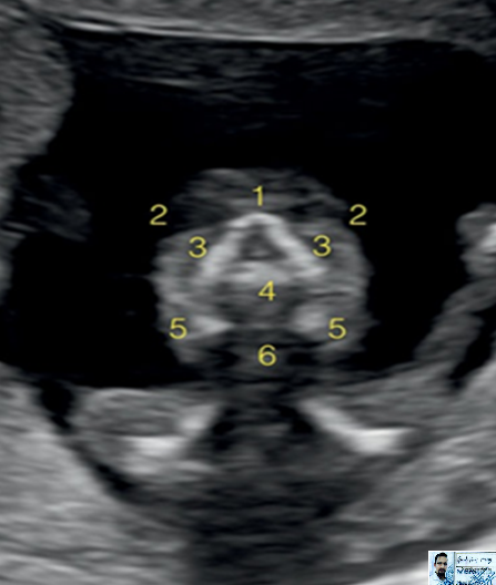

Cranial Structures
1-NT
2-Nasal bone
3-Maxilla
4-Mandible
5-Thalamus
6-Brain stem
7-Fourth ventricle
8-Choroid plexus
9-Cisterna magna


1-Oval head shape
2-Skul bones
3-Falx cerebri
4-Choroid plexus
5-Lateral ventricles
6-Third ventricles
7-Cortex


2-Skul bone
3-Falx cerebri
5-Lateral ventricles
6-Third ventricles
8-Thalimi
9-Aqueduct of sylvius
10-Cerebral peduncles


1- Thalami
2- Developing cerebellum
3- Fourth ventricle
4- Choroid plexus of fourth ventricle
5- Cisterna magna


1- Eyes/Orbit
2- Maxillary processes
3- Nose
3–Facial Structures

At 13 weeks of gestation, the facial structures of a fetus are more defined and continue to develop rapidly.

1-Nasal bone
2-Orbitas/eyes
3-Maxillary processes
4-Maxilla-alveolar ridge
5-Mandible
6-Mandibular gap
4–Neck Structures
Nuchal Translucency
5–Thoracic Structures

1-Lungs
2-Ribs
3-Thorasic aorta
4-Right ventricle
5-Left ventricle


6-Cardiac axis
7-Diastolic ventricular filling


1-Pulmonary
2-Transverse aortic arch
3-superior vena cava
4-Trachea
5-Systolic flow in great vessels

1-Lungs
2-Stomach in abdomen
3-Rib
6–Abdominal Structures

The fetal abdomen should be unobstructed and free from compression by limbs or the umbilical cord.

1-Lungs
2-Stomach in abdomen
3-Rib

6- cord insertion

4- Bladder
5- Two umbilical arteries
6- cord insertion

1-Kidneys
2-Lung
3-Ribs
4-Spine
5-Pelvic bones
7–Limbs and Extremities

Upper and Lower Limbs: Limbs should be positioned to avoid obscuring critical structures, particularly the abdominal wall, face, and spine.

  • Purpose: To assess for limb symmetry and appropriate development.
  • Reference Structures: Long bones (humerus, femur), hands, and feet.
1-Hand and finger
2-Lower arm
3-Upper arm

1-Upper arm
2-Lower arm
3-Hand with fingers

1-Foot
2-Lower leg
3-Upper leg
8–Spine

Spine Alignment: The spine should appear straight or slightly curved, maintaining a natural position without excessive curvature.

  • Purpose: Ensures accurate crown-rump length (CRL) measurement and detection of spinal abnormalities.
  • Reference Structures: Cervical, thoracic, lumbar, and sacral vertebrae.
1-Skin
2-Spine

1-Spine
2-Ribs
3-Scapula

Bilingual Quiz

Note: If you select English, answer all questions in English.
เคฏเคฆि เค†เคช เคนिंเคฆी เคšुเคจเคคे เคนैं, เคคो เคธเคญी เคช्เคฐเคถ्เคจ เคนिंเคฆी เคฎें เคนเคฒ เค•เคฐें।

1. What is the most accurate biometric parameter for dating a pregnancy in the first trimester? 1. เค—เคฐ्เคญाเคตเคธ्เคฅा เค•ी เคคाเคฐीเค– เคจिเคฐ्เคงाเคฐिเคค เค•เคฐเคจे เค•े เคฒिเค เคชเคนเคฒे เคคिเคฎाเคนी เคฎें เคธเคฌเคธे เคธเคŸीเค• เคฌाเคฏोเคฎेเคŸ्เคฐिเค• เคฎाเคช เค•ौเคจ เคธा เคนै?
A. Biparietal diameter (BPD) / เคฌाเค‡เคชेเคฐाเค‡เคŸเคฒ เคกाเคฏเคฎीเคŸเคฐ
B. Femur length (FL) / เคซीเคฎเคฐ เคฒंเคฌाเคˆ
C. Crown-rump length (CRL) / เค•्เคฐाเค‰เคจ-เคฐंเคช เคฒंเคฌाเคˆ
D. Head circumference (HC) / เคธिเคฐ เค•ी เคชเคฐिเคงि
2. What is the typical gestational age range for performing a nuchal translucency (NT) scan? 2. เคจुเค•เคฒ เคŸ्เคฐांเคธเคฒूเคธेंเคธी (NT) เคธ्เค•ैเคจ เค•เคฐเคจे เค•ी เคธाเคฎाเคจ्เคฏ เค—เคฐ्เคญाเคตเคงि เคธीเคฎा เค•्เคฏा เคนै?
A. 8–10 weeks / 8–10 เคธเคช्เคคाเคน
B. 10–12 weeks / 10–12 เคธเคช्เคคाเคน
C. 11–13 weeks + 6 days / 11–13 เคธเคช्เคคाเคน + 6 เคฆिเคจ
D. 14–16 weeks / 14–16 เคธเคช्เคคाเคน
3. An increased nuchal translucency thickness is associated with which condition? 3. เคจुเค•เคฒ เคŸ्เคฐांเคธเคฒूเคธेंเคธी เค•ी เค…เคงिเค• เคฎोเคŸाเคˆ เค•िเคธ เคธ्เคฅिเคคि เคธे เคœुเคก़ी เคนोเคคी เคนै?
A. Macrosomia / เคฎैเค•्เคฐोเคธोเคฎिเคฏा
B. Trisomy 21 / เคŸ्เคฐाเค‡เคธॉเคฎी 21
C. Twin-to-twin transfusion / เคœुเคก़เคตां เคธे เคœुเคก़เคตां เคŸ्เคฐांเคธเคซ्เคฏूเคœเคจ
D. Neural tube defect / เคจ्เคฏूเคฐเคฒ เคŸ्เคฏूเคฌ เคกिเคซेเค•्เคŸ
4. What is the normal upper limit of nuchal translucency (NT) thickness at 12 weeks gestation? 4. 12 เคธเคช्เคคाเคน เค•ी เค—เคฐ्เคญाเคตเคงि เคฎें เคจुเค•เคฒ เคŸ्เคฐांเคธเคฒूเคธेंเคธी เค•ी เคธाเคฎाเคจ्เคฏ เคŠเคชเคฐी เคธीเคฎा เค•्เคฏा เคนै?
A. 2.5 mm
B. 3.0 mm
C. 3.5 mm
D. 4.0 mm
5. What structure should be assessed to exclude acrania or anencephaly in the first trimester scan? 5. เคชเคนเคฒे เคคिเคฎाเคนी เค•े เคธ्เค•ैเคจ เคฎें เคเค•्เคฐेเคจिเคฏा เคฏा เคเคจेเคจเคธेเคซैเคฒी เค•ो เคฌाเคนเคฐ เค•เคฐเคจे เค•े เคฒिเค เค•िเคธ เคธंเคฐเคšเคจा เค•ा เคฎूเคฒ्เคฏांเค•เคจ เค•िเคฏा เคœाเคจा เคšाเคนिเค?
A. Nuchal skin fold / เคจुเค•เคฒ เคค्เคตเคšा เคคเคน
B. Cranial ossification / เค•เคชाเคฒ เค•ी เคนเคก्เคกी เคฌเคจเคจा
C. Amniotic bands / เคเคฎ्เคจिเคฏोเคŸिเค• เคฌैंเคก्เคธ
D. Spine alignment / เคฐीเคข़ เค•ी เคธंเคฐेเค–เคฃ
6. Which of the following is typically not assessed during a routine late first trimester scan? 6. เคจिเคฎ्เคจเคฒिเค–िเคค เคฎें เคธे เค•ौเคจ เค†เคฎเคคौเคฐ เคชเคฐ เคฆेเคฐ เคธे เคชเคนเคฒे เคคिเคฎाเคนी เคธ्เค•ैเคจ เคฎें เค†ंเค•ा เคจเคนीं เคœाเคคा เคนै?
A. Nuchal translucency / เคจुเค•เคฒ เคŸ्เคฐांเคธเคฒूเคธेंเคธी
B. Nasal bone / เคจाเคธिเค•ा เคนเคก्เคกी
C. Uterine artery Doppler / เคฏूเคŸेเคฐिเคจ เค†เคฐ्เคŸเคฐी เคกॉเคชเคฒเคฐ
D. Kidney echogenicity / เค•िเคกเคจी เค•ी เค‡เค•ोเคœेเคจिเคธिเคŸी
7. What is the primary purpose of measuring the nasal bone in the late first trimester scan? 7. เคชเคนเคฒे เคคिเคฎाเคนी เค•े เค…ंเคค เคฎें เคธ्เค•ैเคจ เคฎें เคจाเคธिเค•ा เคนเคก्เคกी เคฎाเคชเคจे เค•ा เคฎुเค–्เคฏ เค‰เคฆ्เคฆेเคถ्เคฏ เค•्เคฏा เคนै?
A. Dating the pregnancy / เค—เคฐ्เคญाเคตเคธ्เคฅा เค•ी เคคाเคฐीเค– เคฒเค—ाเคจा
B. Predicting gestational diabetes / เค—เคฐ्เคญเค•ाเคฒीเคจ เคฎเคงुเคฎेเคน เค•ी เคญเคตिเคท्เคฏเคตाเคฃी
C. Screening for chromosomal anomalies / เค—ुเคฃเคธूเคค्เคฐ เค…เคธाเคฎाเคจ्เคฏเคคाเค“ं เค•ी เคœांเคš
D. Detecting cleft lip / เค• cleft เคนोंเค  เค•ा เคชเคคा เคฒเค—ाเคจा
8. What is a normal fetal heart rate in the late first trimester (11–13 weeks)? 8. เคชเคนเคฒे เคคिเคฎाเคนी เค•े เค…ंเคค (11–13 เคธเคช्เคคाเคน) เคฎें เคญ्เคฐूเคฃ เค•ी เคธाเคฎाเคจ्เคฏ เคนृเคฆเคฏ เค—เคคि เค•्เคฏा เคนोเคคी เคนै?
A. 80–100 bpm
B. 100–120 bpm
C. 120–160 bpm
D. 160–200 bpm
9. In a normal late first trimester scan, what is the status of the yolk sac? 9. เคธाเคฎाเคจ्เคฏ เคฆेเคฐ เคธे เคชเคนเคฒे เคคिเคฎाเคนी เคธ्เค•ैเคจ เคฎें เคฏोเค• เคธैเค• เค•ी เคธ्เคฅिเคคि เค•्เคฏा เคนोเคคी เคนै?
A. Not yet visible / เค…เคญी เคคเค• เคฆिเค–ाเคˆ เคจเคนीं เคฆेเคคा
B. Prominent and pulsatile / เคช्เคฐเคฎुเค– เค”เคฐ เคธ्เคชंเคฆเคจเคถीเคฒ
C. Usually regressing or not seen / เค†เคฎเคคौเคฐ เคชเคฐ เค˜เคŸ เคฐเคนा เคนोเคคा เคนै เคฏा เคจเคนीं เคฆिเค–เคคा
D. Main source of nutrition / เคชोเคทเคฃ เค•ा เคฎुเค–्เคฏ เคธ्เคฐोเคค
10. Which structure is routinely visualized to rule out omphalocele during the late first trimester? 10. เคชเคนเคฒे เคคिเคฎाเคนी เค•े เค…ंเคค เคฎें เค“เคฎ्เคซाเคฒोเคธीเคฒ เค•ो เคฌाเคนเคฐ เค•เคฐเคจे เค•े เคฒिเค เค•िเคธ เคธंเคฐเคšเคจा เค•ो เค†เคฎเคคौเคฐ เคชเคฐ เคฆेเค–ा เคœाเคคा เคนै?
A. Liver echotexture / เคฒिเคตเคฐ เค•ी เค‡เค•ोเคŸेเค•्เคธเคšเคฐ
B. Umbilical cord insertion / เค—เคฐ्เคญเคจाเคฒ เค•ा เคฒเค—ाเคต
C. Amniotic fluid pocket / เคเคฎ्เคจिเคฏोเคŸिเค• เคฆ्เคฐเคต เค•ी เคœेเคฌ
D. Spine / เคฐीเคข़ เค•ी เคนเคก्เคกी

2 comments:

  1. Result / เคชเคฐिเคฃाเคฎ:

    Q1: Correct Answer

    Q2: Correct Answer

    Q3: Correct Answer

    Q4: Correct Answer

    Q5: Incorrect. Correct answer is B

    Q6: Correct Answer

    Q7: Correct Answer

    Q8: Correct Answer

    Q9: Correct Answer

    Q10: Correct Answer

    Your score: 9/10 (90%)

    เค†เคชเค•ा เคธ्เค•ोเคฐ: 9/10 (90%)

    ReplyDelete
  2. My score is 70% sohit kumar from bihar, khagaria

    ReplyDelete

RENAL CALYCEAL MICROLITHIASIS & Prostatomegaly (BPG grade-i) - AN ULTRASOUND DIAGNOSIS whole abdomen male

๐Ÿ–‹️ Genrate reports Fig-1. rENAL shows   Single tiny echogenic focus (~2 mm)  noted in mid calyx  without definite posterior acoustic shado...

Popular post