Lobes of the Liver
The liver is divided into anatomical and functional lobes, based on surface landmarks and internal vasculature respectively.
| Lobe |
Location |
Sonographic Landmarks |
| Right Lobe |
Largest; right of IVC and GB fossa |
Bordered medially by MHV and GB |
| Left Lobe |
Left of falciform ligament |
Extends across midline |
| Caudate Lobe |
Posterior to porta hepatis, between IVC & ligamentum venosum |
Seen posteriorly in transverse scan |
| Quadrate Lobe |
Inferior and anterior, between GB and ligamentum teres |
Often considered part of left lobe |
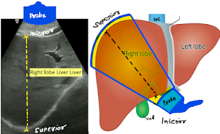
1. Right lobe:
The right lobe of the liver is the largest lobe, located on the right side of the body, extending from the midline to the right abdominal wall. It lies to the right of the gallbladder fossa and inferior vena cava (IVC). It is separated from the left lobe by the middle hepatic vein (MHV) and the main lobar fissure.
Key Sonographic Points:
1. Largest of the liver lobes
2. Located right of the IVC and gallbladder
3. Borders
Medially: Middle hepatic vein, gallbladder
Superiorly: Diaphragm
Posteriorly: Right kidney
4. Best visualized in longitudinal and transverse subcostal views.
2. Left lobe:
The left lobe of the liver is the second largest lobe, located to the left of the falciform ligament, and often extends across the midline into the left upper quadrant of the abdomen. It lies anterior to the stomach and superior to the pancreas.
Key Sonographic Points:
1. Lies left of the falciform ligament, often extending over the stomach and pancreas.
2. Separated from the right lobe by the falciform ligament and middle hepatic vein (MHV)
3. Inferior border aligns with the ligamentum teres
4. Ultrasound Appearance
Appears anterior to the aorta, celiac axis, and pancreas
Seen clearly in epigastric and subxiphoid views
Sometimes appears elongated or enlarged, especially in thin individuals
5. Important site for left lobe masses, focal fatty changes, or metastatic deposits
6. Crosses the midline, making it easily visible in both transverse and longitudinal scans
3. Caudate lobe:
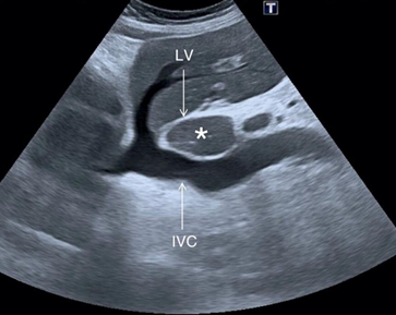
The caudate lobe is a small, anatomically distinct lobe of the liver located on the posterior surface, nestled between the inferior vena cava (IVC) and the ligamentum venosum. Despite its small size, it has separate vascular supply and venous drainage, making it functionally significant.
Key Sonographic Points:
1. Posterior to the porta hepatis
2. Bounded by the IVC (right side) and ligamentum venosum (left side)
3. Lies superior to the caudate process, above the porta hepatis
4. Borders:
Right: IVC
Left: Ligamentum venosum
Inferior: Porta hepatis
Anterior: Posterior segment of the left lobe
5. Vascular Supply:
Receives blood from both right and left portal veins
Has direct venous drainage into the IVC through short hepatic veins
Ultrasound Appearance:
Seen posterior to the liver hilum in a transverse epigastric scan
May appear prominent in cases of liver cirrhosis (caudate hypertrophy)
Key landmark in evaluating portal hypertension
4. Quadrate lobe:
The quadrate lobe is a small glandular segment of the liver situated on the inferior anterior surface. It lies between the gallbladder fossa on the right and the ligamentum teres on the left, and is functionally considered by some as part of the left liver.
Couinaud Segmentation: Used for surgical and imaging localization, dividing the liver into eight functionally independent segments, each with its own portal vein, hepatic artery, and bile duct branch.
Key Sonographic Points:
1. Location: Positioned on the visceral (inferior) surface, nestled between the gallbladder and the ligamentum teres.
2. Boundaries:
Medial/Right: Gallbladder fossa
Medial/Left: Ligamentum teres (round ligament)
Anterior/Inferior: Liver diaphragmatic surface
Posterior: Porta hepatis and MHV influence
3. Sonographic Characteristics
Visualized primarily in epigastric transverse scans, best seen through subcostal or epigastric windows just above the gallbladder.


No comments:
Post a Comment