Anatomy of the Liver-Liver Ultrasound Perspective

Topic Banner
ANATOMY OF THE LIVER- Liver Ultrasound Perspective

Liver Lobes and Segments


1. Lobes:
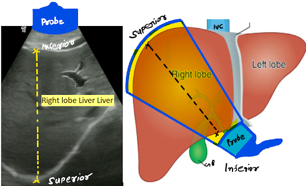
Right lobe: Largest; located in the right hypochondrium

Left lobe: Extends into the epigastric region

Caudate lobe: Posterior to the left lobe, adjacent to the inferior vena cava (IVC)

Quadrate lobe: Medial segment of the left lobe, near the gallbladder

Couinaud Segmentation: Used for surgical and imaging localization, dividing the liver into eight functionally independent segments, each with its own portal vein, hepatic artery, and bile duct branch.

2. Couinaud Segmental Anatomy (8 Segments)

Liver Segments and Landmarks

Liver Segments and Main Landmarks

Segment Location Main Landmarks
I Caudate lobe Posterior to portal vein, near IVC
II Left superior lateral Above left portal vein
III Left inferior lateral Below left portal vein
IVa/b Left medial (superior/inferior) Medial to falciform ligament
V Right anterior inferior Below right portal vein, anterior
VI Right posterior inferior Below right portal vein, posterior
VII Right posterior superior Above right portal vein, posterior
VIII Right anterior superior Above right portal vein, anterior

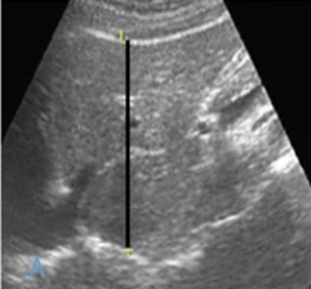
3. Diaphragm thickness
The normal thickness of the diaphragm around the liver, measured by ultrasound, is between 2.2mm -2.8mm. centimeters in healthy volunteers. A diaphragm thickness of less than 0.2 centimeters at the end of exhalation is considered a sign of diaphragm atrophy.
Thickening fraction is calculated according to the formula = (EIT − EET)/EET × 100.

Topic Banner
ANATOMY OF THE LIVER- Liver Ultrasound Perspective

Vascular Anatomy


Portal Vein System
  • Formed by the confluence of the superior mesenteric and splenic veins
  • Has echogenic walls on ultrasound due to collagenous content
  • Enters the liver at the porta hepatis, dividing into right and left branches
  • Supplies ~75% of liver’s blood flow
The portal venous system can be recognised on ultrasound as a tubular structure with echogenic walls that enters the liver together with the hepatic artery (HA) at the level of the hepatic hilum (a), and reaches the more distal liver segments. (b) Posterior branch of the right portal vein (RPV); (c) left portal vein (LPV) branches. (c) The caudate lobe can be clearly visualised in this scanning plane (asterisk) between the inferior vena cava (IVC), the ligamentum venosum (LV), and LPV. CBD, common bile duct; EHPV, extrahepatic portal vein.
Hepatic Artery
  • Arises from the celiac trunk
  • Supplies ~25% of liver’s blood
  • Usually seen adjacent to the bile duct and portal vein (the "portal triad")

Hepatic Veins
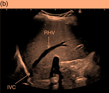
  • Right, middle, and left hepatic veins drain into the IVC
  • No echogenic walls; typically seen as anechoic channels converging superiorly
The hepatic veins originate from the periphery of the liver, converging into the inferior vena cava (IVC). LHV, left hepatic vein; MHV, middle hepatic vein; RHV, right hepatic vein. Topic Banner
ANATOMY OF THE LIVER- Liver Ultrasound Perspective

Biliary System


Intrahepatic Bile Ducts
  • Run alongside portal vein branches
  • Not normally visible unless dilated
The common bile duct (CBD) can be seen as a thin tubular structure with echogenic walls that, in the majority of cases, runs anteriorly and parallel to the portal vein (PV) at the level of the hepatic hilum. The hepatic artery (HA) is often seen at this level in transverse section, hence it is visualised as a small rounded or ovoid structure (depending on the angle of insonation) with echogenic walls between the CBD and the PV.
Extrahepatic Biliary System
  • Includes the common hepatic duct, cystic duct, and common bile duct (CBD)
  • CBD is often evaluated in the porta hepatis region
  • Normal CBD diameter: ≤ 6 mm in adults (can increase with age or post-cholecystectomy)
The gallbladder (GB)-
The gallbladder (GB) is a pear‐shaped structure located in the GB fossa, a depression on the visceral surface of the liver between the right and left lobe. The GB is usually lateral to the second part of the duodenum and anterior to the right kidney (RK). (b) Note is made of the main interlobar fissure (IF) between the portal vein (PV) and the GB.
Topic Banner
ANATOMY OF THE LIVER- Liver Ultrasound Perspective

Ligaments and Landmarks


Falciform Ligament
  • Divides left and right lobes anteriorly
  • Sometimes visible as a thin echogenic line on ultrasound
Ligamentum Teres (Round Ligament)
  • Remnant of fetal umbilical vein
  • Seen as an echogenic focus within the left lobe
Ligamentum teres (LT) or round ligament takes direct contact with the left branch of the portal vein (LPV). LT runs along the ventral surface of the liver continuing with the falciform ligament (FL) along the dorsal surface of the liver. By changing the plane of insonation from transverse to longitudinal scan view (left to right image) the LT will be seen elongating in full extent to join the FL (right side image).
Ligamentum Venosum
  • Remnant of fetal ductus venosus
  • Separates the caudate lobe from the left lobe
Ligamentum Venosum
  • Remnant of fetal ductus venosus
  • Separates the caudate lobe from the left lobe
The boundaries of the caudate lobe
The boundaries of the caudate lobe (asterisk) are defined by the retrohepatic inferior vena cava (IVC), the ligamentum venosum (LV), and the left branch of the portal vein (LPV) that is better seen when imaging in transverse section (left side image).

No comments:

Post a Comment

Umbilical-urachal sinus with preperitoneal abscess SCRS

116 Case Study Umbilical-urachal sinus with preperitoneal abscess. An umbilical-urachal sinus is a congenital anomaly r...

Popular post